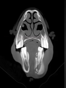
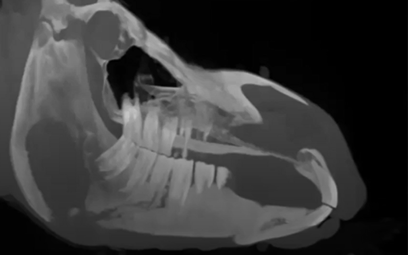
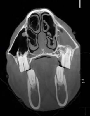
This is a 9 yo hunter jumper that had the 10 tooth extracted and developed an oral sinus fistula. A plug was inserted, meanwhile CT images were taken, and a tract was shown running into her sinus. A protemp bridge was inserted (prosthetic tooth), the bone filled in and healed well. Featured: Dr. Travis Henry, University of Wisconsin-Madison
Read MoreThis was an older mare that sustained trauma to her mandible in the pasture. CT images show a draining tract and sequestrum, the tooth above the draining track was removed.
Featured: Dr. Travis Henry, University of Wisconsin-Madison
Read MoreThis horse had advanced TMJ pathology, which caused mastication issues. CT images show compression on the temporal hyoid bones and guttural pouch.
Featured: Dr. Travis Henry, University of Wisconsin-Madison
Read MoreThis Warmblood mare came in with sinusitis. CT images show a Diastema which was the cause of the sinusitis. The 10 tooth was removed and Protemp (human crown) was applied to create a prosthetic tooth.
Featured: Dr. Travis Henry, University of Wisconsin-Madison
Read MoreThis horse has all the clinical symptoms of periodontal disease. CT images show an increase in periodontal ligament width, horizontal/vertical bone loss, periodontal infection, diastema, food impaction causing infection to spread and creation of a draining tract.
Featured: Travis Henry, University of Wisconsin-Madison
Read MoreThis middle aged QH presented nasal discharge, believed to be sinusitis. Upon CT scans dense dental material was found in the nasal passage. This material was causing the nasal discharge.
Featured: Dr. Travis Henry, University of Wisconsin-Madison
Read MoreThis 14 yo Percheron had plenty of dental issues. CT images show several missing teeth, expanded mandibles, a previous mandibular fracture, a severe shear mouth, and fluid in the sinuses.
Featured: JR Lund, University of Wisconsin-Madison
Read MoreThis 18 yo TB cross had a persistent draining tract and bone sequestrum. The mare had the affected area surgically debrided. The majority of the bone sequestrum was removed. Plus an additional case study at the end.
Featured: Dr. JR Lund - University of Wisconsin
Read MoreThis is a 13 yo Belgian who had a history of nasal discharge and a previous tooth fracture. CT Images show a palatable crown fracture and secondary sinusitis.
Featured: Dr. Jr Lund-University of Wisconsin Madison
Read MoreThis is a 10 yo Quarter Horse with a two week history of head shaking mostly on the right side. This horse received a CT scan, osteo-cyst lesions and sclerosis were found on the TMJ and were treated with injections.
Featured: Dr. Jr Lund-University of Wisconsin Madison
Read More


