Horley, UK – Asto CT Inc. is excited to announce the continued growth of their Equina standing, computed tomography (CT) scanner in the UK market.
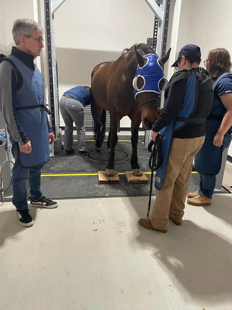
Read More“The Equina® is one of the most innovative, advanced imaging devices available for veterinary medicine. We can scan front or back limb pairs, or the head/neck of a standing horse under light sedation, allowing the patient to be in and out in just minutes with minimal stress and risk.”
Read More"The trouble is, of course, you can't see it; these horses (at risk) look fine, they won't give any hint there's a problem lurking there. So, that's why we need some form of screening. Imaging with CT is probably what's required to get the images to show the trainer."
Read More”A major advantage of the Equina standing CT scanner is that it images limb pairs. This allows the horse to stand naturally with minimal positioning involved. Veterinarians can then compare both limbs [affected and unaffected] to find the cause for lameness.”
Read MoreOne of the leading private equine practices in Texas, Equine Sports Medicine & Surgery (ESMS), has installed the first equine standing CT (Computed Tomography) scanner in the state. ESMS, which has one of the highest volumes of equine cases in the United States, is excited to add the Asto CT Equina® scanner to their full-service suite of equine veterinary services to improve patient outcomes and welfare.
Read MoreOn top of that, there were no true equine CT scanners on the market, and most clinicians had to be content with using what amounted to a human machine that was repurposed for equine use. But what if there was a better way? Why settle for only ‘good enough’?
Read MoreAsto CT makers of the Equina, is proud to announce the release of its 92cm Tapered Bore with extended cervical spine and proximal limb coverage.
Read MoreWe went through our archives to display the five most common limb injuries and diseases identified by the Equina. The 1st most common limb issue identified by the Equina is Navicular Syndrome….
Read MoreRead more about Dianne's story and knowledge with equine diagnostic imaging.
“If you can get diagnostic imaging that gives you the answer quickly, and safely, it’s a huge advantage. That’s why I’m such an advocate for the Equina by Asto CT, because it would have given us the information we needed in 30 seconds compared to hours.”
Read MoreImaging with computed tomography (CT) has become increasingly advanced in recent years and has become the gold standard diagnostic for dental disease and other conditions involving the skull.
Read MoreWe would like to officially welcome Dr. Sabrina Brounts to the Asto CT team as a scientific advisor.
Read More“Hoof abscesses occur when bacteria get trapped between the sensitive laminae (the tissue layer that bonds the hoof capsule to the coffin bone) and the hoof wall or sole. The bacteria create exudate (pus), which builds up and creates pressure behind the hoof wall or sole. This pressure can become extremely painful.” -Dr. Brian Fitzgerald
Read MoreWhen a horse goes under general anesthesia, there is an inevitable risk, as there is evidence of a link between increased equine mortality and general anesthesia. This causes many horse owners to feel uneasy about the procedure. All the information poses the ultimate question, how do you take a CT scan of a massive animal without administering general anesthesia?
Read More


